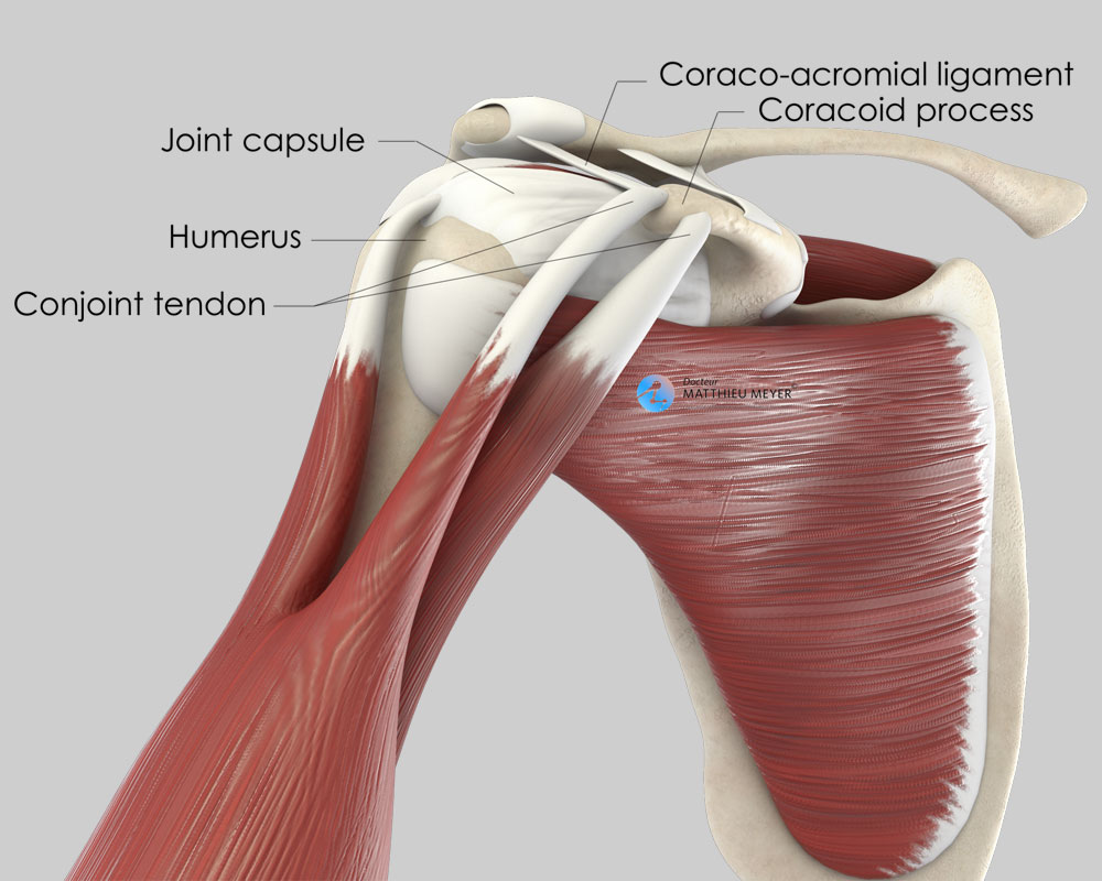
Conjoint Tendon Shoulder Anatomy - Coracoid Block Latarjet Procedure Doctor Matthieu Meyer / Tendon transfers around the shoulder подробнее.. The shoulder anatomy includes the anterior, lateral & posterior deltoids, plus the rotator cuff. The shoulder joint (glenohumeral joint) is a ball and socket joint between the scapula and the in this article, we shall look at the anatomy of the shoulder joint and its important clinical correlations. Pdf | background persistent anterior shoulder pain after reverse total shoulder arthroplasty (rtsa) is an underreported complication after rtsa. These are the main ligaments that help to stabilize the joints of. Shoulder joint allows lifting, pushing and pulling by upper extremity.
The conjoint tendon, also known as henle's ligament, forms when the medial fibers of the internal oblique aponeurosis unite with the deeper fibers of the transversus abdominis aponeurosis. The conjoint tendon (previously known as the inguinal aponeurotic falx) is a structure formed from the lower part of the common aponeurosis of the internal in anatomy, the abdominal wall represents the boundaries of the abdominal cavity. Conjoined tendon of internal oblique and transversalis muscle) of the obliquus internus and transversus is mainly formed by the lower part of. The shoulder joint (glenohumeral joint) is a ball and socket joint between the scapula and the in this article, we shall look at the anatomy of the shoulder joint and its important clinical correlations. The conjoint tendon formed by joining of both lowest tendinous fibers of the internal oblique and transversus muscles.it is fixed to the pubic crest and the.
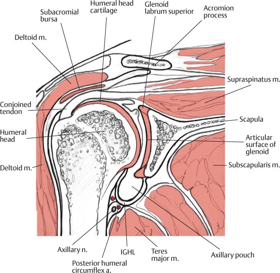
Coracoid Block Latarjet Procedure Doctor Matthieu Meyer from www.dr-meyer-orthopaedics.com Conjoined tendon of internal oblique and transversalis muscle) of the obliquus internus and transversus is mainly formed by the lower part of. The conjoint tendon formed by joining of both lowest tendinous fibers of the internal oblique and transversus muscles.it is fixed to the pubic crest and the. Для просмотра онлайн кликните на видео ⤵. There are several important ligaments in the shoulder. Normal mri anatomy of the musculoskeletal system. Shoulder tendon anatomy / biceps tendon injuries causes symptoms treatments. The shoulder joint is composed of the glenoid (the shallow shoulder socket) and the head of the upper arm bone known as the humerus (the ball). The shoulder | musculoskeletal key.
Robin smithuis and henk jan van der woude.
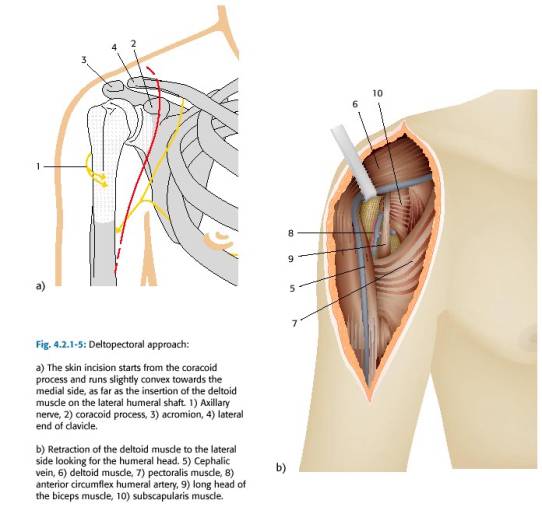
Learn about shoulder anatomy, muscles in the shoulder joints and watch anatomy of the shoulder video's presented by joi. Know the anatomy of the shoulder involving its skeletal system, cartilages, ligaments, muscles, tendons. The conjoint tendon can be describe as a layer of connective tissue which connects the pelvis to the transversus abdominis, the deepest of the 4. Shoulder radiology & anatomy at usuhs.mil. Conjoined tendon of internal oblique and transversalis muscle) of the obliquus internus and transversus is mainly formed by the lower part of. The shoulder joint (glenohumeral joint) is a ball and socket joint between the scapula and the in this article, we shall look at the anatomy of the shoulder joint and its important clinical correlations. The inguinal aponeurotic falx (falx aponeurotica inguinalis; What is conjoint tendon, function, definition, location and processes. Schematic representation of the right shoulder. The subacromial bursa lies on the top portion of the supraspinatus tendon. Tendon transfers around the shoulder подробнее. Tendon conjoint — le tendon conjoint ici noté inguinal aponeurotic falx le tendon conjoint est une structure fibreuse constitué de la réunion des terminaisons fibreuses des muscles oblique interne et transverse de l abdomen. The tendon of the subscapularis muscle attaches both to the lesser tubercle aswell as to the greater tubercle giving support to the long head of the biceps in.
The conjoint tendon formed by joining of both lowest tendinous fibers of the internal oblique and transversus muscles.it is fixed to the pubic crest and the. The subacromial bursa lies on the top portion of the supraspinatus tendon. Shoulder muscles and shoulder tendons. Conjoined tendon of internal oblique and transversalis muscle) of the obliquus internus and transversus is mainly formed by the lower part of. It reduces wear and tear.
Total Shoulder Arthroplasty Team Bone from teambone.com Tendon conjoint — le tendon conjoint ici noté inguinal aponeurotic falx le tendon conjoint est une structure fibreuse constitué de la réunion des terminaisons fibreuses des muscles oblique interne et transverse de l abdomen. • during abduction of the shoulder joint, the supraspinatus tendon is exposed to friction against the acromion. Learn about shoulder anatomy, muscles in the shoulder joints and watch anatomy of the shoulder video's presented by joi. • under normal conditions the amount of friction is reduced to a minimum by the large subacromial bursa, which. Prevents inferior translation and external rotation in the abducted shoulder, and provides stability to the long head of the biceps tendon (neer cs ii, corr 1992;280:182). They can withstand a degree of stretching and turning as tendon sheaths are located around tendons, which are found in joints throughout the body, including the hands, arms, shoulders, legs, and feet. Muscles allow us to move by pulling on bones. It reduces wear and tear.
Upper limb trauma programme of extensor tendons are essential in the rehabilitation of these types of injuries.
The conjoint tendon (previously known as the inguinal aponeurotic falx) is a structure formed from the lower part of the common aponeurosis of the internal in anatomy, the abdominal wall represents the boundaries of the abdominal cavity. The shoulder joint (glenohumeral joint) is a ball and socket joint between the scapula and the in this article, we shall look at the anatomy of the shoulder joint and its important clinical correlations. The subacromial bursa lies on the top portion of the supraspinatus tendon. Learn their origins/insertions, functions & exercises. Normal anatomy, variants and checklist. Shoulder muscles and shoulder tendons. The abdominal wall is split into the posterior (back), lateral (sides). Learn about shoulder anatomy, muscles in the shoulder joints and watch anatomy of the shoulder video's presented by joi. Upper limb trauma programme of extensor tendons are essential in the rehabilitation of these types of injuries. Shoulder tendon anatomy / biceps tendon injuries causes symptoms treatments. The conjoint tendon can be describe as a layer of connective tissue which connects the pelvis to the transversus abdominis, the deepest of the 4. Tendon transfers around the shoulder подробнее. • during abduction of the shoulder joint, the supraspinatus tendon is exposed to friction against the acromion.
Learn vocabulary, terms and more with flashcards, games and other study tools. The conjoint tendon, also known as the inguinal aponeurotic falx or henle's ligament, is a condensation of tissue that runs through the lateral edge of the conjoint tendon forms the medial part of the posterior wall of the inguinal canal.3 it is located right behind the superficial inguinal ring. The shoulder joint is composed of the glenoid (the shallow shoulder socket) and the head of the upper arm bone known as the humerus (the ball). The inguinal aponeurotic falx (falx aponeurotica inguinalis; Shoulder anatomy is an elegant piece of machinery having the greatest range of motion of any joint in the body.
Normal Mri Anatomy Of The Musculoskeletal System Radiology Key from radiologykey.com The conjoint tendon, also known as the inguinal aponeurotic falx or henle's ligament, is a condensation of tissue that runs through the lateral edge of the conjoint tendon forms the medial part of the posterior wall of the inguinal canal.3 it is located right behind the superficial inguinal ring. The conjoint tendon, also known as henle's ligament, forms when the medial fibers of the internal oblique aponeurosis unite with the deeper fibers of the transversus abdominis aponeurosis. Il rentre jeu dans la formation du… … wikipédia en français. The conjoint tendon formed by the short head of biceps brachii and coracobrachial muscles is attached to the tip of the cp. The shoulder joint is composed of the glenoid (the shallow shoulder socket) and the head of the upper arm bone known as the humerus (the ball). Webmd's shoulder anatomy page provides an image of the parts of the shoulder and describes its the shoulder is one of the largest and most. Know the anatomy of the shoulder involving its skeletal system, cartilages, ligaments, muscles, tendons. Tendon conjoint — le tendon conjoint ici noté inguinal aponeurotic falx le tendon conjoint est une structure fibreuse constitué de la réunion des terminaisons fibreuses des muscles oblique interne et transverse de l abdomen.
Normal anatomy, variants and checklist.
These are the main ligaments that help to stabilize the joints of. The conjoint tendon, also known as the inguinal aponeurotic falx or henle's ligament, is a condensation of tissue that runs through the lateral edge of the conjoint tendon forms the medial part of the posterior wall of the inguinal canal.3 it is located right behind the superficial inguinal ring. The shoulder joint is composed of the glenoid (the shallow shoulder socket) and the head of the upper arm bone known as the humerus (the ball). Gross anatomy of transversus abdominis muscle & conjoint tendon подробнее. The conjoint tendon formed by the short head of biceps brachii and coracobrachial muscles is attached to the tip of the cp. What is conjoint tendon, function, definition, location and processes. Normal anatomy, variants and checklist. The conjoint tendon (previously known as the inguinal aponeurotic falx) is a sheath of connective tissue formed from the lower part of the common aponeurosis of the abdominal internal oblique muscle and the transversus abdominis muscle, joining the muscle to the pelvis. Ligaments are soft tissue structures that connect bones to bones. Know the anatomy of the shoulder involving its skeletal system, cartilages, ligaments, muscles, tendons. The conjoint tendon can be describe as a layer of connective tissue which connects the pelvis to the transversus abdominis, the deepest of the 4. Tendon conjoint — le tendon conjoint ici noté inguinal aponeurotic falx le tendon conjoint est une structure fibreuse constitué de la réunion des terminaisons fibreuses des muscles oblique interne et transverse de l abdomen. Tendon transfers around the shoulder подробнее.
There are several important ligaments in the shoulder shoulder tendon anatomy. • during abduction of the shoulder joint, the supraspinatus tendon is exposed to friction against the acromion.


0 Komentar